


February 13, 2020 by John Fernandez
3D Printing Helps Improve Patients’ Cardiac and Vascular Care

When Mel Dick, age 80, had a minimally invasive procedure to treat an aneurysm in his left leg earlier this year, he never thought he’d return to Miami Cardiac & Vascular Institute just a few months later for an additional intervention.
The reason for the second interventional procedure was uncovered by his doctor, Barry Katzen, M.D., during a routine exam in Mr. Dick’s hospital room. Dr. Katzen, founder and chief medical executive of Miami Cardiac & Vascular Institute, revealed a second popliteal aneurysm – this time behind Mr. Dick’s right knee.
Video by Steve Pipho
(VIDEO: Barry Katzen, M.D., founder and chief medical executive of Miami Cardiac & Vascular Institute, uses 3D printing to create models of patients’ anatomy for use in interventional procedures, surgeries and patient education.)
A popliteal aneurysm is a bulging in the artery that supplies blood to the knee, thigh and calf. This type of aneurysm occurs more often in men. For Mr. Dick, it meant the aneurysm in his right knee would need to be repaired soon after he recovered from the procedure performed on his left knee.
“I never expected it,” said Mr. Dick of Sunny Isles, Fla. “I’m so thankful he [Dr. Katzen] found it. If not, I could have lost my leg – or worse.”
If a popliteal aneurysm is left untreated, a blood clot can form and cut off blood supply to the leg, often leading to amputation. And if the aneurysm were to burst, life-threatening bleeding can occur. When diagnosed early, a minimally invasive procedure can prevent either of these things from happening.
To help plan the procedure and explain it to his patient, Dr. Katzen used emerging technology to create a 3D model of Mr. Dick’s knee joint. With a few clicks on a computer keyboard, Dr. Katzen selected CT scan images taken of the knee and transmitted them to a special printing lab.
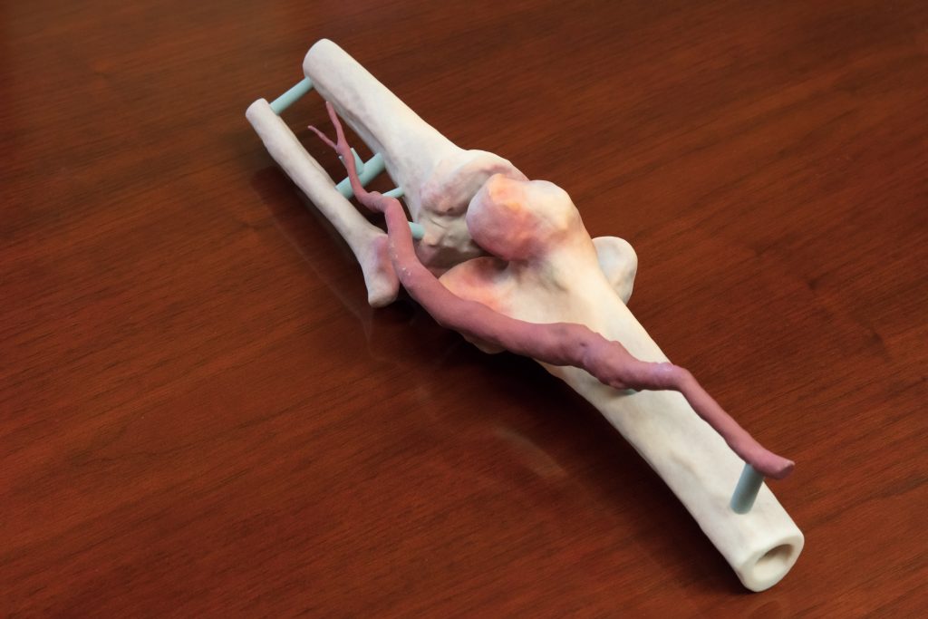
There, 3D printing was used to create a life-size, anatomically correct model of Mr. Dick’s knee. The model of the knee joint, made out of calcium carbonate, feels like actual bone and includes the aneurysm in real size, as well as the surrounding anatomy.
“The patient is literally holding their body part in their hands,” Dr Katzen said. “We’re taking CT scan data and translating it into ways that can help us treat patients. The 3D model also has a practical effect in helping us learn the patient’s anatomy. We often bring the model into the procedure room to use as a guide and reference.”
Dr. Katzen says the 3D model helps him and his medical team be more precise while carefully navigating around delicate veins and arteries. The 3D printing benefits patients by seeing what will be repaired, helping them to better understand the problem and the procedure that will take place to fix it.
“This is wonderful,” Mr. Dick said. “Now I understand exactly what’s going to happen, and I can go home and explain it to my family.”
The Baptist Health News Team was there to capture the first time Dr. Katzen presented a 3D model to his patient. Watch the video to now to see the patient’s reaction and learn more about how 3D printing is helping to advance medicine.
top stories

Technology 
There are no comments